docvbansal@gmail.com
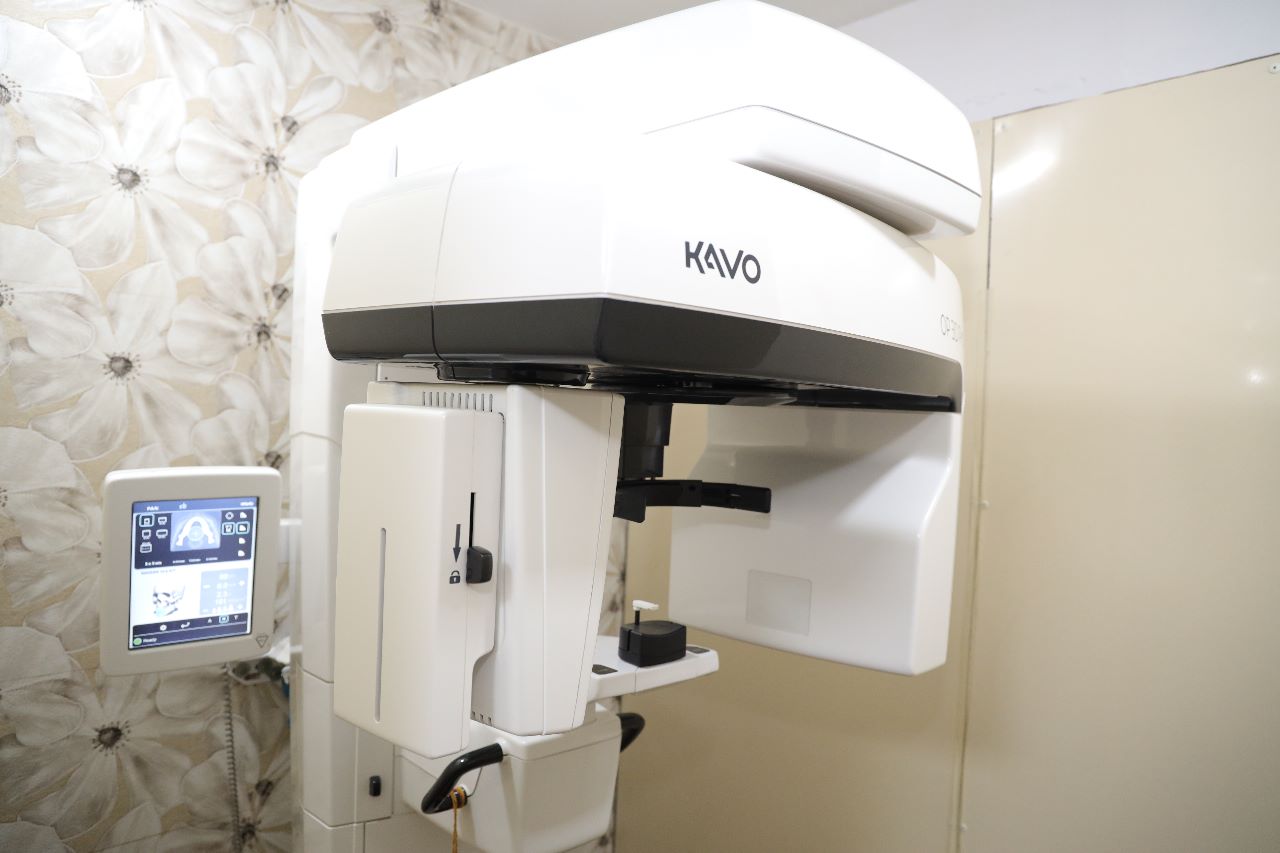
CBCT is the most exciting advance in dental imaging which has revolutionized the field of diagnostic radiology. Being a 3 d imaging modality it makes the radiographic image crystal clear and there is little scope for doubt, misjudgment of misinterpretation (associated with conventional radiographs).
Bansal Dental Care is one of the few dental centers in india equipped with cbct machines which enables perfect pretreatment planning & post surgical evaluation. This also gives flexibility of checking in between procedures.
CBCT technology is being widely used in all fields of dentistry, including orthodontics, endodontics, oral surgery/pathology, periodontics, and implant treatment planning.
A conventional ct scan, or “Computed Tomography” is a sort of medical imaging that combines several x-ray readings into virtual “slices” of an object, allowing the physician to look within the object without cutting into it.
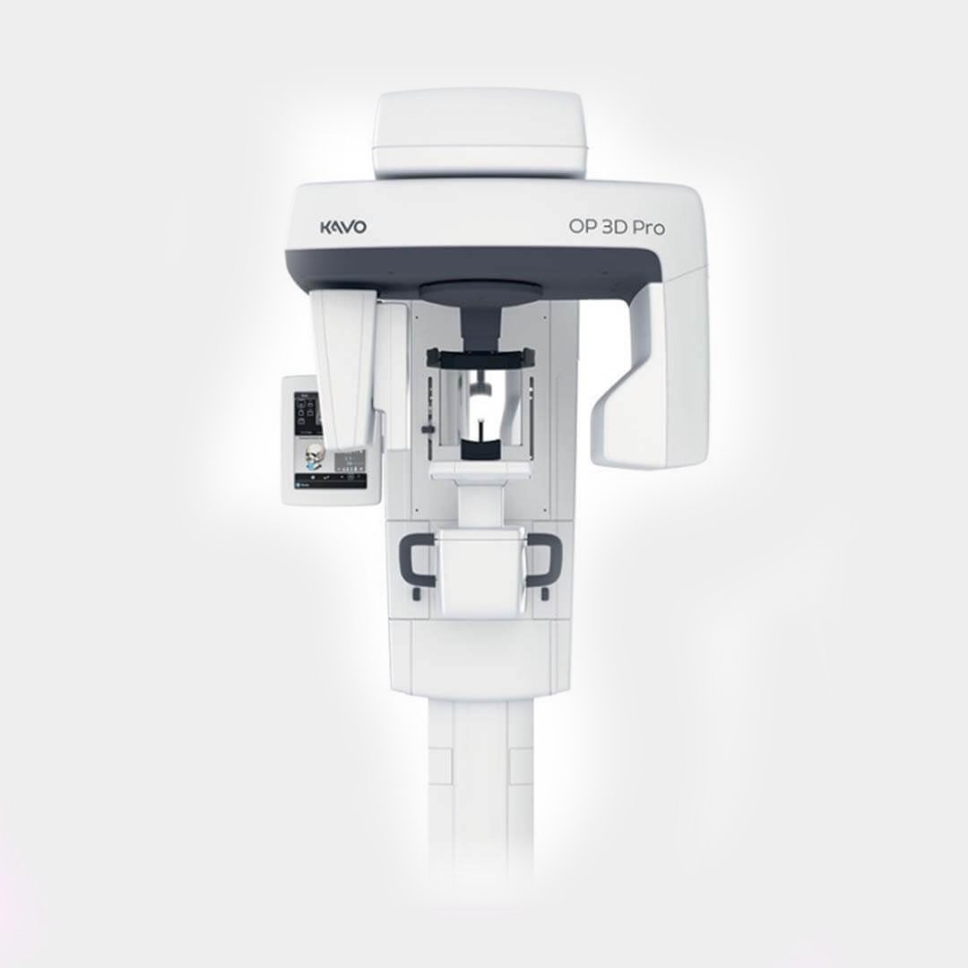
A sub-type of computed tomography known as cone-beam computed tomography (CBCT) is used in dentistry, orthodontics, oral surgery, and endodontics.
Today, we will look a little deeper into this fascinating technology that is increasingly getting crucial in identifying and treating dental problems.
Technology is continuously developing and evolving. At Bansal Dental Care, we make every effort to stay updated with our patient’s needs by utilizing sophisticated technologies.
To ensure a high success rate and give you the best dental implant, we generate a 3d model of your mouth from which a digital blueprint for your dental implants is created. We have an in-house 3D x-ray facility, known as a cone beam computed tomography (CBCT) scan, that allows us to precisely diagnose all problems and devise treatment strategies.
Dr. Vineet Bansal , one of the best dentists in yamunanagar, Haryana utilizes CBCT as the first step to ensure that your implants fit precisely with maximum customization and precision.
Dental cone beam computed tomography is an x-ray machine utilized when ordinary dental or face X-rays aren’t enough. This ct scanner uses specific techniques to create 3-d images of dental structures, nerve routes, soft tissues, and bone in the craniofacial region in a single scan.
Since the radiation dose from this scanner is substantially higher than that of typical dental X-rays, dentists rarely utilize it regularly. More detailed treatment planning is possible thanks to images generated using cone-beam CT.
An OPT (Orthopantomogram) is a panoramic scanning dental x-ray of the upper and lower jaw. It is also sometimes called by the proprietary name orthopantomography or Ranorex. It shows a flattened two-dimensional view of a half-circle from ear to ear. Panoramic X-rays allow images of multiple angles to be taken to make up the composite panoramic image, where the maxilla (Upper Jaw) and mandible (Lower Jaw) are in the viewed area. The structures that are outside the viewed area are blurred. At some stage in your dental treatment, your dentist will likely take an OPT. An OPT also demonstrates the number, position, and growth of all the teeth including those that have not yet surfaced or erupted through the gum. It is different from the small close-up X-rays dentists take of individual teeth. It shows less fine detail, but a much broader area of view. This can be particularly useful to check hard-to-see areas like wisdom teeth, or the development of a child’s jaw and teeth. It is also often used to check your jaw joint, the TMJ (temporomandibular joint), sometimes called the CMA (craniomandibular articulation), especially if you grind your teeth.
Your dentist will ask you to remove any jewelry or metallic items from around the head and neck area as it can interfere with the image, blocking the x-ray. The OPT usually takes about 3 mins to complete, only 20 seconds of this are the image being taken.
Bansal Dental Care has an opg machine on site for your convenience and the images are produced digitally and stored in your file with all your other X-rays. The image is produced within seconds of the opg being taken and your dentist will review it with you at the end of the appointment. This can be as part of a dental check up, Or separate appointment. An opg is just one diagnostic tool your dentist will use to ensure you maintain and prevent unnecessary dental issues.
RVG or Radiovisiography is the most recent technique of imaging used in dentistry. The radiation exposure is minimal as compared to the other traditional techniques used for capturing images.
RVG stands for Radiovisiography and is a technology that has replaced traditional X-ray radiology methods. It is used extensively in dentistry now due to the benefits that it is to offer.
Bansal Dental Care is equipped with two rvg. We are using rvg since 2006. RVG is a integral part of our practice.